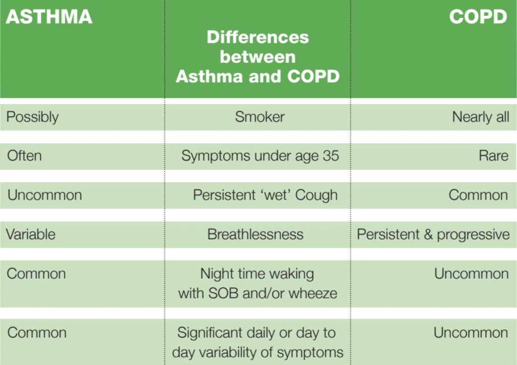
Asthma is a common, longterm disease that requires ongoing management. If you have asthma, you have very sensitive airways – the tubes that carry air in and out of your lungs. Certain triggers can cause your airways to become inflamed and tighten when you breathe. Triggers can include stress, exercise, cold air, and breathing in particular substances such as smoke, pollution or pollen. (British Lung Foundation definition)
Key facts
• 5.4 million people in the UK are currently receiving treatment for asthma: 1.1million children (1 in 11) and 4.3 million adults (1 in 12).
• In 2016 1,410 people in the UK died from asthma.
• Asthma attacks hospitalise someone every 8 minutes; 185 people in the UK are admitted to hospital because of asthma every day (a child is admitted every 20 minutes)

The anatomy of an asthmatic attack
Whilst everyone’s airways will react to inhaled irritants, asthmatic patients, due to the underlying low-grade inflammation, over-react. An asthmatic attack occurs in two stages. Initially there is an early asthmatic response characterised by a fall in peak flow rate due to bronchoconstriction. This will resolve within about 3 hours. In atopic individuals excess immunoglobulin E (IgE) is produced and attaches to mast cells. Interaction will allergens cause de-granulation of the mast cells leading to the release of a range of bronchospastic mediators including; histamine, bradykinin and leukotrienes and this results in a late asthmatic response that can last for 12 to 16 hours.
The chronic inflammatory responseresults in characteristic changes;
• Airway epithelium may be shed
• Excess mucus secretion
• Mediators affect autonomic control of airways resulting in bronchoconstriction

Management of Asthma
Non-drug
Smoking cessation will improve the management of asthma. The benefits of taking exercise, eating a healthy diet and correcting obesity are evident. Overweight/obese patients will suffer greater morbidity from their asthma. Avoiding allergens (where possible) will help.
Drug Management
Drug management of asthma can be spilt into two therapeutic groups; bronchodilators (relievers) and antiinflammatory agents (preventers).
Bronchodilators
Beta-2-adrenoreceptors are found within the bronchial smooth muscle. Beta-2 agonists interact with these receptors resulting in bronchial smooth muscle relaxation. In addition, they inhibit mediator release from mast cells and increase mucociliary clearance.
• Short acting(SABA) Salbutamol, Terbutaline
• Long-acting(LABA) Salmeterol, Formoterol
Due to their rapid, almost immediate, onset of action short-acting beta-2- agonists are mostly administered by inhalation as a dry powder (DPI) or a metered dose inhaler (MDI). Beta-2-agonists are not fully selective for beta-2-receptors and can affect beta-1-receptors resulting in the common side-effects, including; tremor, cramps, palpitations, tachycardia, hypokalaemia and peripheral vasodilation.
The long-acting beta-2-agonists have a duration of action of approximately 12-15 hours. They are not intended for relief of acute symptoms but symptom prophylaxis. They are therefore indicated for the control of nocturnal asthma and to prevent exercise induced bronchospasm.
Anticholinergics (antimuscurinic bronchodilators -LAMA)
• Tiotropum
Anticholinergic agents competitively inhibit the action of acetylcholine at
the muscurinic receptors resulting in bronchodilation. Tiotropium is now licensed for the treatment of Asthma; it is not appropriate for the relief of acute bronchospasm.
These drugs should be used with caution in patients suffering from glaucoma, prostatic hyperplasia and bladder outflow obstruction. Sideeffects include dry mouth, nausea, constipation and headache.
Methyxanthines
• Theophylline
These drugs are not effective when administered by inhalation. They have a complex and uncertain mode of action but are effective bronchodilators licensed for the management of asthma. They relaxsmooth muscle, inhibit mediator release, increase mucociliary transport and suppress oedema.
Theophylline has a narrow therapeutic window (5-20 micrograms/ml) -great care must be taken administering the drug concomitantly with drugs known to induce/inhibit drug metabolism. Common drugs that raise plasma
theophylline levels include; cimetidine, ciprofloxacin, clarithromycin, the
COC pill and diltiazem. Drugs that lower theophylline levels include; carbamazapine, phenobarbitone, phenytoin, rifampacin and cigarette smoke.
Side-effects associated with theophylline and might be associated with toxicity include; nausea and vomiting, abdominal discomfort, CNS stimulation and sleeplessness. At higher plasma levels (above 35 micrograms/ml) SE’s are potentially lethal including; arrythymias, palpitation, tachycardia and convulsions.
Anti-inflammatory agents
• Corticosteroids-ICS
• Beclomethasone
• Fluticisone
• Mometasone
• Prednisolone• Compound preparations ICS/LABA
• Budesonide/Formoterol
• Fluticasone/Formoterol
• Fluticasone/Salmeterol
• Fluticasone/Vilanterol
• Beclomethasone/formoterol
Corticosteroids suppress and inhibit all elements of the inflammatory response; they stop mediators being released from inflammatory cells and suppress the activity of these mediators.
They are particularly effective in the late asthmatic response and have
been demonstrated to;
• Have an anti-inflammatory effect on the bronchial mucosa
• Reduce airways hyper-responsiveness
• Reduce bronchial oedema.
SE’s of the drug, especially following chronic use, are greatly reduced by use of the inhaled route.
These include growth suppression in children, adrenal suppression and
effect on bone metabolism.
Leukotriene receptor antagonists (LTRA)
• Montelukast
• Zafirlukast
These drugs act by blocking the effects of leukotriene (mediators) in the airways and thus have an antiinflammatory effect. They are not for use in acute attacks.
Montelukast has not been shown to be more effective compared to corticosteroids but the two drugs appear to have an additive effect.
SE’s include GI disturbances, dry mouth, thirst and hypersensitivity reactions as well as CNS effects. CSM has advised that patients prescribed LTRAs should be alert to the development of eosinophilia, vasculitic rash, worsening pulmonary symptoms, cardiac complication and peripheral neuropathy.

BTS/SIGN Guidance on Management of Asthma in Adult
It is recommended to start at a step appropriate to initial severity of the condition.
Step 1. Infrequent short lived wheeze
Inhaled short-acting bronchodilator, as required.
Consider monitored initiation of treatment with low dose ICS
Move to step 2 (if uncontrolled)
Step 2. Regular Prevention Inhaled short-acting bronchodilator, as required. Start Inhaled Corticosteroid at low dose Move to step 3 (if uncontrolled)
Step 3. Initial add on therapy
Add inhaled LABA to low dose ICS (usually a combination inhaler)
Move to step 4 (if uncontrolled) Step 4. Additional add-on therapies If no response to LABA-stop LABA and consider increase dose ICS If benefit to LABA but control still inadequate -continue LABA and increase ICS to medium dose If benefit to LABA but control still inadequate -continue LABA and ICS and consider addition of LTRA, SR Theophyline or LAMA Move to step 5 (if uncontrolled)
Step 5. High -dose therapies
Consider trials of:
• increasing ICS to high dose
• Addition of 4th Agent e.g. LTRA, SR Theophyline, Beta agonist tablet, LAMA
Refer Patient for specialist care
(*only consider step up once compliance has been assured and correct inhaler technique verified)
Stepping down should only occur where the patient has been stable for 3 months. Rescue oral steroids can be used at any step to manage severe asthma exacerbations.
Drug delivery and Inhaler Technique
Not all inhalers are equal, there are two specific types:
1.Metered Dosage Inhalers (MDI’s) including Soft Mist Inhaler, and Breath activated versions including K-haler, Easi-Breathe and Autohalers.
2.Dry powder inhalers (DPI’s) which are essentially all breath activated devices (BA).
The inhalation techniques/Inspiratory effort needed for each are
very different:
• MDI-Slow, steady and deep (Aerosol made by device) Inspiratory effort needed is low (optimally 30l/min)
• DPI-Hard, fast and deep (patient generates the aerosol by using
their breath to aggregate the drug particles to a small enough size to reach the lower parts of the lung).
Inspiratory effort needed depends on the device (optimally 60-
90l/min.) Inspiratory flow is checked using an In-Check Dial device.
A spacer device should be used with MDIs to co-ordinate actuation and promote better lung drug delivery whilst reducing the oral deposition of drug.
The breathe actuated MDI devices should be considered when inhalation technique demonstrated is slow and steady but the co-ordination of inspiration and depressing the canister is poor. These automatically release the aerosol when the inspiration reaches the optimum level.
Compliance with the inhaler and dosing regimen are critical aspects of achieving maximal therapeutic outcomes.
The patients’ ability to use any device is a key aspect of inhaler selection. Manual dexterity, understanding of how to use the device, along with knowledge of why they need to use are also of significant importance.
Nebulisers are no more efficient than an MDI device used with a spacer (10 separate puffs delivered over 5-10 minutes) but for some patients, those with only shallow breathing, there might be benefit.

